Giant Bedroom
Thoracic surgeon Errol Bush discusses what surgical options are available for lung cancer patients, the recovery and aesthetic benefits of minimally invasive surgery for lung cancer, and when it's appropriate and why it's important to be treated at a comprehensive cancer center, such as the Johns Hopkins Lung Cancer Program.
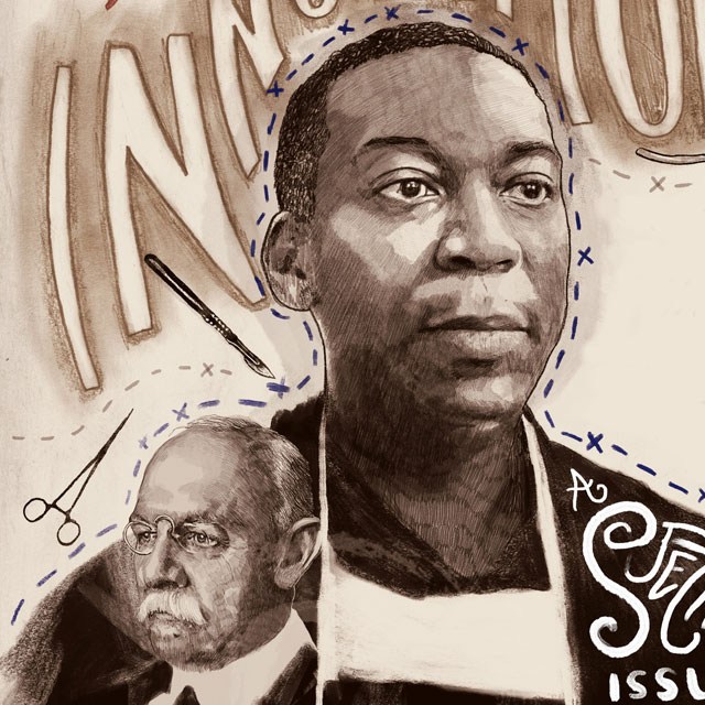
Innovators in Surgery
William Halsted & Errol Bush
Nineteenth-century surgeries were harrowing experiences: no cocktails of anesthetics to numb the pain, no antibiotics to prevent infection, no procedures in place to prevent excessive bleeding. Remarkably, all that changed within a few decades thanks to the systematic training of physicians and the rapid dissemination of revolutionary ideas. Key to that revolution was William Stewart Halsted, whose two years of training in Europe endowed him with an understanding of germ theory and German surgical instruction.
During his time at Johns Hopkins (1886–1922), Halsted furthered his earlier experiments with cocaine as an anesthetic and developed techniques for intestinal and stomach surgeries, hernia and aneurysm repair, and thyroid operations. Most famously, he was the first in the United States to perform a radical mastectomy to treat breast cancer, pioneering a procedure that now bears his name (the Halsted radical mastectomy).
The standout surgeon is also renowned for his vigilant control of bleeding, obsession with cleanliness and gentle handling of patient tissue. But all of those advances may have remained within the walls of The Johns Hopkins Hospital had he not understood the importance of medical education. Halsted did everything surrounded by medical students and residents, whose training he helped to formalize. In this way, "Halsted's principles" were passed on to the next generation of surgeons, who instilled them in the surgery departments at medical institutions throughout the country.
Inheriting this legacy is Errol Bush, the surgical director of the advanced lung diseases and lung transplant program at Johns Hopkins. Currently, Bush is the only surgeon at Johns Hopkins dedicated to performing lung transplants. Of the 50 he has performed in his two years here, not a single patient has died.
Bush also does his best during operations to avoid cardiopulmonary bypass (CPB), which involves rerouting a patient's blood to a device outside the body to obtain oxygen and release carbon dioxide. CPB adversely affects outcomes for some patients. But for those patients who do need CPB (about 40 percent of the total, including those with weak hearts and those unable to survive on a single lung), Bush likes to use a newer, "lighter" version of CPB. It requires less of the anticoagulant heparin, resulting in less bleeding and less inflammation, and therefore improved outcomes.
In addition, Bush is currently spearheading Johns Hopkins' participation in a multicenter, international trial to test an investigational "lung-in-a-box" device that could significantly expand the pool of donor lungs. The technique, called ex vivo lung perfusion, circulates a blood-based preservation solution through the lungs to keep them fresh for four to 10 hours while they are treated with antibiotics and cleaned of secretions and debris that may have accumulated. If inflammation and infections clear up sufficiently and the lungs can be used, another transplant recipient gets to leave the waiting list.
"Fifteen to 30 percent of patients die before new lungs become available, so every lung we salvage is a life that is saved," says Bush. Halsted would be proud.
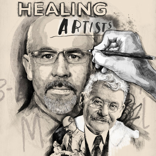
Healing Artists
Max Brödel & Juan Garcia
Joining the residents who peered over the shoulders of William Stewart Halsted during operations was a young artist from Germany named Max Brödel. Through his lifelike illustrations, he shared his operating table-side view with physicians around the world, giving them detailed insights into the pioneering techniques of Halsted, gynecologist Howard Kelly and other Johns Hopkins legends.
Brödel first worked with Carl Ludwig, a well-known German physician who needed help conveying the physiological research he was doing in Leipzig. That collaboration turned Brödel into a scientist and de facto medical student. He never touched pencil to paper until he had a clear understanding of his subject, often going to great lengths to do so. Gary Lees, recently retired chair of the Department of Art as Applied to Medicine, founded by Brödel in 1911 as the nation's first such department, notes that the illustrations that came from Brödel's studies pushed medicine forward by giving hundreds of physicians a view inside the body, helping them plan their procedures.
Today, Juan García is doing the same as director of the 3-D Printing and Visualization Facility at the Carnegie Center for Surgical Innovation. There, instead of using the ink and carbon dust that Brödel employed, he utilizes ultraviolet light-cured plastic resin of different transparencies, colors and flexibilities to transform two-dimensional patient images—like CT and MRI scans—into 3-D models that surgeons can hold in their hands. In so doing, García equips doctors with patient-specific anatomical details.
García's most challenging project yet was for a patient with severe scoliosis of the spine. Special software helped him convert static CT images into a 3-D model of the S-shaped spine, a task that required two print jobs totaling 69 hours, plus hours more to assemble the delicate jigsaw puzzle. The resulting model will allow spine surgeons to preoperatively plan incisions and placement of corrective hardware to decrease the surgery's time and improve results.
García also cares for patients directly at the Johns Hopkins Facial Prosthetics Clinic. There, he uses his background as a painter and sculptor to design "wearable medical art"—silicone prostheses for patients who have lost part of their face to cancer or trauma. "These are not for cosmetic purposes," says García. "They restore a patient's sense of wholeness, allow them to reintegrate into society, provide functional support for eyeglasses and protect the sensitive cavities left by their surgeries."
García begins with scans or impressions of the patient's face and then sculpts or 3-D prints a model for the silicone prosthesis so that it will be exactly the right shape. Then, he painstakingly paints the prosthesis to match the patient's skin tone and features, making his final touches with the prosthesis in place. Just as Brödel's work was not art for art's sake, neither is García's. This is life-changing rehabilitative medical art.
Brainstem Pathfinders
Florence Sabin & Sascha du Lac
Sascha du Lac had been at Johns Hopkins for two years before she got to know Florence Sabin, Class of 1900. Du Lac's colleague, Elisabeth Glowatski, had just been named the first female full professor of the Department of Otolaryngology–Head and Neck Surgery (and the school of medicine's 213th)—which made du Lac wonder who had been the school's first. That's when Google helped du Lac learn more about the influential scientist whose work ultimately earned her a place in the U.S. Capitol's Statuary Hall.
Sabin came to Johns Hopkins as a medical student in 1896 but soon realized that she preferred the peace and quiet of the laboratory to the busyness of the clinic. One of her first projects was to study the brainstem of infants by drawing detailed pictures of 48 tissue sections cut in two planes. To accurately capture the tiny bundles of nerve fibers, she used a "camera lucida" to project an enlarged image of the tissue onto a translucent piece of frosted glass for tracing. She then converted those tracings into wax plates to reconstruct a 3-D model of the entire structure, which Max Brödel illustrated. Together, their work formed Sabin's book An Atlas of the Medulla and Midbrain, published in 1901. It was the first to reveal the neural connections between the brainstem and the spinal cord, and it quickly became a standard textbook in the field.
Sabin went on to elucidate the embryonic development of the lymphatic system, blood vessels, blood cells and connective tissue. She was also a dedicated mentor and teacher. In 1925, she moved to the Rockefeller Institute for Medical Research to pursue research full time.
In contrast, du Lac came to Johns Hopkins in 2013 from an exclusive research institution to be immersed in a clinical environment. At the Salk Institute, she had studied the vestibulo-ocular reflex, the automatic movements our eyes make to counteract our head movements and keep our sight from getting blurry. These movements are initiated by the brainstem, which plays an integral role in balance, among other things.
Her scientific research became personal when her mother was diagnosed with lung cancer and chronic obstructive pulmonary disease. du Lac noticed that when lack of oxygen caused her mom's balance to be off, her cognitive abilities were low and her anxiety was up, but when her balance was good, so were her memory, reasoning and mood. Was there a connection? "That question compelled me to seek answers," she says. "And I realized that a lot of what we know as neuroscientists hasn't made it into clinical practice yet."
Now du Lac spends as much time as possible in the clinic so that she can focus her research on more clinically relevant problems. While attending rounds, she has been able to suggest diagnoses for a few patients based on her intimate knowledge of the brainstem in mice. Her latest work uses genetic techniques to "color-code" specific types of neurons in the brainstem to discover their connections or to make subsets of neurons responsive to light so that they can be selectively stimulated to see their effects.
With today's technology, du Lac is continuing Sabin's work of clarifying the anatomy of the brainstem, going beyond structure to understand function.
Trailblazers in Diabetes
John Jacob Abel & Gerald Hart
Diabetes is among the oldest diseases ever described, first appearing in an Egyptian manuscript from 1552 B.C. But it wasn't until about 1889 that the medical community began to understand it as a problem of the pancreas. Eventually, the hypothesis was posited that diabetes results from a deficiency of a substance, dubbed insulin, secreted by specific pancreatic cells. Finally, in 1921, scientists demonstrated the reversal of diabetes in dogs by giving them an extract—dubbed insulin—from specific pancreatic cells of healthy dogs. But no one knew what the substance of that extract was.
Enter renowned biological chemist John Jacob Abel, who joined the Johns Hopkins University School of Medicine at its inception in 1893 as director of the Department of Pharmacology and Physiological Chemistry. He was well-known for his chemical purification of hormones, particularly that of epinephrine/adrenalin, from the adrenal glands of cows. He had also created a primitive dialysis machine, used in laboratory animals.
The purification of insulin came in 1927, when Abel was 70. After more than three years of trying, he was finally greeted by beautiful crystals of insulin growing on the sides of a test tube. Further tests suggested that insulin was a protein, which countered the assumptions of his day. This information helped other researchers understand insulin's regulation and, eventually, locate its gene and clone it into bacteria to mass produce it.
Today, still tackling the problem of diabetes is Gerald Hart, chair of what now is called the Department of Biological Chemistry. Hart's contributions revolve around a sugar called O-linked N-acetylglucosamine, or O-GlcNAc, which he discovered in 1983. Instead of acting as an energy source, this sugar acts as a thermostat for energy availability. When glucose abounds, so does O-GlcNAc, which gets attached to all sorts of proteins, modifying their behavior. When Hart first discovered this phenomenon, textbooks claimed that sugar modification—or glycosylation—only occurs on cell surface proteins and secreted proteins. Hart showed that in actuality, most proteins in the interior of the cell are glycosylated.
Gradually, the link between chronic high blood sugar and diabetes is becoming clear. When O-GlcNAc levels remain too high, many cellular processes stop working well, Hart says. Sustained over a long period of time, these subtle changes can have dire consequences, so it isn't too surprising that O-GlcNAc has also been linked to diseases like cancer and Alzheimer's.
In addition to altering mitochondrial enzymes that affect cellular metabolism, chronically high levels of O-GlcNAc alter gene activity. Hart's most recent work shows that the addition of O-GlcNAc to the protein TBP changes the activity of more than 400 genes that all relate to cellular energy and lipid metabolism.
While finding a drug to appropriately alter O-GlcNAc's levels throughout the body is a daunting task, Hart has hope scientists will find something that works. In the meantime, he has encouraging results for detecting prediabetes with a simple blood test, which could help people get their sugar levels under control before any serious damage is done.


Source: https://www.hopkinsmedicine.org/news/publications/hopkins_medicine_magazine/features/on-the-shoulders-of-giants
 Reviewed by terrie
on
Desember 01, 2021
Rating:
Reviewed by terrie
on
Desember 01, 2021
Rating:


Tidak ada komentar: